Three-dimensional anatomy and dynamic changes of the conjunctival fornix assessed by high-resolution magnetic resonance imaging
To the best of our knowledge, this is the first study to characterise the 3D anatomy of the conjunctival fornix and its dynamic changes in different eye positions. By quantifying FD, C-F distance and IFS volume and surface area across the primary gaze, upward gaze, downward gaze and eyelid closure, we provide novel insights into a region that has traditionally been regarded as a static space with little clinical significance. This paradigm shift from a static to a dynamic anatomical concept has important implications for both clinical evaluation and device design.
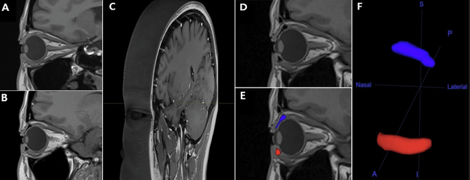
Despite the insertion of sponges with identical volumes, the superior IFS demonstrated a larger volume and surface area than the inferior IFS. This suggests that the superior fornix may exhibit greater compliance and flexibility, thereby serving as a principal tear reservoir [7]. In a disease status, superior fornix involvement would likely lead to more pronounced clinical consequences and render it a critical structure for reconstructive surgery [5,6,7, 20]. From a therapeutic perspective, the superior fornix may also provide more favourable site than the inferior fornix for accommodating implants of a given size. Moreover, because IFS volume and surface area did not differ significantly across gaze positions in our dataset, these findings suggest that physiologic eye movements alone may not impose sufficient deformation to measurably compress a forniceal insert.
Notably, FD and C-F distance exhibited gaze-dependent variability, reflecting the dynamic nature of the fornix. The superior FD was shortest during downgaze and significantly increased during upgaze and eyelid closure. The inferior FD was shortest at primary gaze and significantly increased during eyelid closure. These findings suggest that the positional behaviour of fornix-based implants may vary depending on eye position and eyelid state [17]. Similarly, the superior C-F distance was shortest during eyelid closure, likely reflecting the combined effects of lid closure and Bell’s phenomenon, which brings the fornix apex closer to the corneal surface. This finding suggests a potential risk of closer implant-cornea proximity during eyelid closure, which may be relevant during sleep [21, 22]. In contrast, the inferior C-F distance was longest at primary gaze and shortest at downgaze, further highlighting the mechanical interplay between the globe and eyelids. Collectively, these results may provide preliminary framework for assessing device safety with respect to eye position and for guiding surgical strategies in fornix reconstruction.
Clinically, diseases that shorten the fornix, such as ocular cicatricial pemphigoid and symblepharon, have been graded primarily on slit-lamp based assessments of horizontal involvement [23, 24]. More recent grading systems, such as that proposed by Kheirkhah and colleagues, incorporate fornix length and width to guide surgical planning and graft sizing, leading to improved outcomes [25]. However, these approaches remain restricted to planar or subjective evaluations. Our study demonstrates that MRI-based quantification provides an objective, reproducible and 3D assessment of fornix anatomy, while capturing dynamic changes not detectable with conventional methods [9, 18, 26, 27]. As the fornix plays a key role in tear distribution and ocular motility, improved understanding of its dynamics may enhance surgical strategies and guide the design of fornix-based devices [1, 6, 11].
Unlike our expectations, in our MRI-based study, the mean superior FD at primary gaze was 14.2 mm, which was shorter than those reported in FDM-based studies: 15.3 mm in healthy South Asians and 15.6 mm in healthy White Caucasians [9, 18]. Similarly, the inferior FD in our study (7.4 mm) was shorter compared to those reported by Khan et al. and Jutley et al. (both 10.9 mm) [9, 18]. These discrepancies may be attributed to methodological differences. In our study, the inserted buckling sponge expanded the fornix but did not increase its anatomical length. In contrast, the FDM may have increased the measured FD by mechanically stretching the fornix, as the device is often pushed all the way to the innermost corner of the fornix by the examiner during use. Moreover, FD measurements using the FDM may be overestimated, as the device must be slightly lifted to avoid corneal contact and to ensure clear visibility of the scale.
Populations or sex specific anatomic variations could also be a contributing factor, as previous normative studies with South Asian, White Caucasian, Turkish and Asian Chinese populations consistently reported that conjunctival fornix depth decreases with age and is generally smaller in females than males [9, 18, 26, 27]. (Supplementary Tables 1, 2) Ethnic variation, likely linked to craniofacial and orbital morphology, has been proposed, but absolute fornix depth values appear broadly comparable across populations. Recent Chinese data by Tan et al. also reported no significant differences when compared with Caucasian, South Asian and Turkish datasets [9, 18, 26, 27]. Thus, age and sex seem to be stronger determinants of FD than ethnicity, although population-specific normative datasets remain important for accurate interpretation.
In this study, all participants were Korean women in their 40 s, resulting in reduced cohort heterogeneity but limiting generalisability. Future large-scale normative studies including broader age ranges and both sexes of South Korean ethnicity would be helpful. Nevertheless, because age-related reductions in fornix depth are more pronounced after 50–60 years, the gaze-dependent patterns observed in this cohort may still provide meaningful reference data [9, 18, 26, 27].
Beyond demonstrating the feasibility of MRI-based assessment of fornix dynamics, this study suggests several directions for technical development. The imaging workflow and segmentation process could be further refined and potentially automated. While fully automated fornix quantification is not yet available, recent MRI studies of adjacent ocular structures—such as the eyeball, optic nerve and orbit—show that semi-automated morphometric extraction is achievable [28,29,30]. These precedents, together with our findings, suggest the possibility of automated fornix-specific morphometry, particularly with high-resolution ocular MRI and standardised gaze registration. However, given the thin and deformable nature of the fornix, a semi-automated landmark- or contour-based approach may be a more realistic initial step. In addition, the development and validation of such methods will require larger multicentre datasets to improve robustness, clinical applicability and generalisability.
Furthermore, MRI-derived anatomical data may be integrated with computational modelling to improve the design of fornix-based implants and to predict implant-fornix interactions. In ophthalmology, finite element and computational fluid dynamics approaches have already been used to connect anatomy, mechanics and device behaviour. For example, dynamic MRI has been combined with finite element modelling to estimate optic nerve head strain during eye movements [31,32,33,34,35]. In the implant field, computational modelling of glaucoma drainage devices has demonstrated that geometry, flow resistance, insertion angle and tissue interaction significantly influence device performance [36, 37]. These precedents suggest that future fornix imaging could be used not only descriptively, but also predictively. Patient-specific MRI-derived fornix geometry may provide the basis for finite element or fluid-structure models of implant-fornix interaction in the future. Such models could help optimise implant design, identify configurations prone to focal compression or instability and improve preclinical evaluation before broader clinical application.
Our study has several limitations. The small sample size, along with the inclusion of a single ethnicity and sex cohort, limits the generalisability of the findings. Additionally, sponge insertion, though necessary for MRI visualisation, may have altered the native fornical configuration; its stiffness, shape and compressibility could have affected expansion patterns. Thus, the present results likely represent sponge-expanded rather than purely native anatomy. Future studies involving larger and more diverse populations, imaging approaches that reduce or avoid artificial expansion, will be important to validate and extend these findings.
In conclusion, this proof-of-concept study demonstrates gaze-dependent forniceal configuration and that high-resolution MRI can be used to quantify its anatomy. Although the findings should be interpreted in the context of a small cohort and sponge-expanded anatomy, they provide a basis for future studies of fornix morphology and for imaging-based evaluation of diseased fornix and fornix-targeted devices.